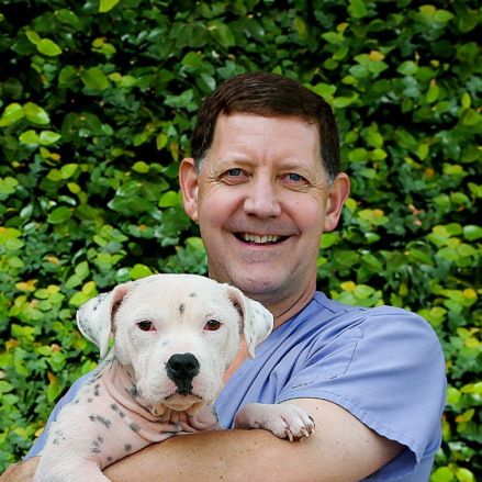
Dr Ryan is interested in 3D training models that are useful for learning techniques. For example, coiling vessels to treat a bleeding sinonasal tumour in a horse, guttural pouch empyema in the horse, intrahepatic porstosystemic shunts in the dog; and use of subcutaneous ureteral bypass in cats with ureteral stones.
Dr. Stewart Ryan, BVSc(Hons), MS is Senior Lecturer and Head of Small Animal Surgery service at UVet veterinary Hospital, Faculty of Veterinary and Agricultural Sciences at The University of Melbourne.
1. Briefly tell us about your research/projects.
As a clinician researcher, most of my research focus areas are related to questions we encounter in the clinic. I have been developing the interventional radiology (IR) service at the University of Melbourne UVet Hospital in Werribee. Interventional radiology utilises fluoroscopy to visualise wires and catheters and stents that are placed in blood vessels or other luminal structures (e.g. urethra, trachea or colon). Some of my other research areas are in the fields of musculoskeletal oncology, the interaction of normal and neoplastic bone with radiation, cartilage and bone healing and surgical oncology.
Intrahepatic portosystemic shunts (IHPSS) are congenital vascular anomalies that allow blood pass directly from the portal vein to the caudal vena cava, bypassing the liver. Open surgical approaches work well for extrahepatic portosystemic shunts but correction of intrahepatic shunts are associated with a high morbidity and mortality rate. An endovascular approach through the jugular vein uses fluoroscopy to place a stent in the caudal vena cava and then thrombogenic coils in the abnormal shunt vessels. To date, seven cases have been treated this way and outcome has been very excellent. As with any new procedure, there is a steep learning curve. I and working with a final year DVM student to develop a 3D vascular model of the three most common shunt morphologies utilising CT angiograms that are acquired during the planning phases of these procedures to practice this procedure.
2. Who are your research colleagues?
I have several collaborative relationships with medical physicians based at St. Vincent’s Hospital, Colorado State University, RMIT and Wollongong University. I have been fortunate to do some preliminary work with colleagues at the Australian Synchrotron evaluating microbeam radiation therapy for treatment of osteosarcoma.
3. What imaging equipment are you using?
Currently, we are using a Phillips BV fluoroscopy image intensifier unit at the Werribee VBIC node. This equipment is quite old and is nearing its end of life. Unfortunately, we do not have digital subtraction angiography (DSA) as part of our current fluoroscopy system, which is particularly advantageous in endovascular IR cases. We are keen to purchase a new fluoroscopy unit with DSA capability that can be used in IR-related research projects.
4. How did you find accessing and using the equipment (through VBIC)?
Being part of the VBIC node at Werribee, it is very useful to have access to the variety of imaging equipment that can be used for research. I regularly use the 16 slice CT unit and 1.5T MRI unit in both clinical and research applications.
5. Thank you Dr Ryan!


0 Comments