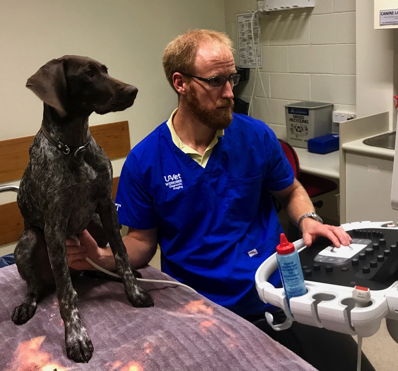
This month we feature research from Robert Turner of Werribee node. Robert’s research work is attempting to establish whether a relationship exists between body composition and fat distribution in dogs and the appearance of their pancreas on ultrasonography and computed tomography.
Briefly tell us about your research project.
My project is attempting to establish whether a relationship exists between body composition and fat distribution in dogs and the appearance of their pancreas on ultrasonography and computed tomography.
In human medicine, it has been shown that an increase in body fat, especially abdominal visceral fat, is related to fat deposition in the pancreas (pancreatic steatosis) which can be seen on ultrasound and CT. Pancreatic steatosis has been associated with the development of a variety of metabolic diseases such as diabetes and pancreatitis. This concept of visceral fat distribution and deposition into organs, such as the pancreas, has had limited research in dogs.
With this in mind, I am hoping to develop a means to non-invasively quantify the abdominal visceral fat in dogs and extending this methodology into assessing for a relationship between fat distribution and the appearance of the pancreas. This may offer a useful model for future investigations into visceral fat distribution and diseases in dogs such as diabetes mellitus, cardiovascular disease and pancreatitis.
Who are your supervisors/colleague/research team members?
Associate Professor Caroline Mansfield is my primary supervisor who is a renown researcher in veterinary gastroenterology with a particular interest in pancreatitis, inflammatory bowel disease and colitis. My co-supervisors include Dr Dayle Tyrrell, the Head of Diagnostic Imaging at the U-Vet Werribee Animal Hospital, and Professor Frank Dunshea, a well respected and leading expert in nutrition and growth physiology. I am also working in affiliation with Cameron Nowell at Monash Institute of Pharmaceutical Sciences who is offering his expertise in image analysis.
What imaging equipment are you using?
I am using a Philips EPIQ5 ultrasound machine with the 5-18MHz linear-array transducer or 3-8MHz convex-array transducer, a Siemens Somatom Emotion 16 Excel Edition CT and a Hologic Discovery QDR Series Dual-energy X-ray Absorptiometry (DXA) machine.
How did you find accessing and using the equipment?
The use of the ultrasound and CT is a continuation of my current residency in Diagnostic Imaging Department at the U-Vet Werribee Animal Hospital. The access and technical support to the equipment are facilitated by the Diagnostic Imaging Department and their radiographers (Kane Wilson and Sylvia Meekings). The DXA machine is provided by Frank Dunshea’s research team, with training and assistance from Evan Bittner and Marree Cox’s.
Did you work across sites? If so, how did you find accessing the equipment across sites?
The diagnostic imaging is being performed on site at the U-Vet Werribee Animal Hospital. Input into imaging analysis is being provided by Cameron Nowell at Monash Institute of Pharmaceutical Sciences.


0 Comments