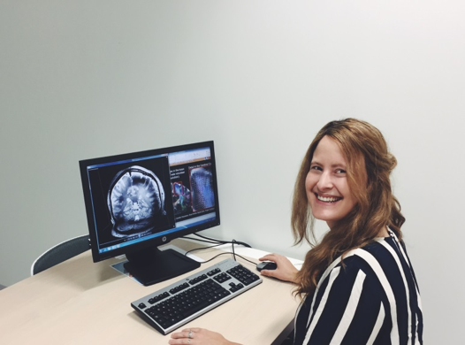
This month we feature research from Myrte Strik of Parkville node. Myrte’s Ph.D. work focuses on the integration of gait biomechanics and 7 Tesla MRI to monitor and predict the progression of gait dysfunction in multiple sclerosis (MS) patients.
Briefly tell us about your research project.
My name is Myrte Strik and I am a jointly awarded PhD student in the Department of Anatomy and Neuroscience at the University of Melbourne, Australia and the VU University Amsterdam, the Netherlands. The aim of my PhD project is to integrate gait biomechanics and 7 Tesla MRI to monitor and predict the progression of gait dysfunction in multiple sclerosis (MS) patients. I am specifically interested in investigating the subtle changes in walking in early stage MS using functional and structural network imaging.
Who are your supervisors?
I am lucky to have not one but four supervisors, Dr. Scott Kolbe and Dr. Jon Cleary in Melbourne and Professor Dr. Jeroen Geurts and Dr. Menno Schoonheim in the Netherlands. Dr. Scott Kolbe is a research fellow in the Department of Anatomy and Neuroscience at the University of Melbourne. Scott’s research has focused on developing MRI techniques to study the structure and function of the brain and how it changes in people with brain disease. Dr. Jon Cleary is a University of Melbourne McKenzie Fellow and has a MBBS-PhD from University College London. His research focuses on developing clinical applications for 7T MRI. In the Netherlands, Jeroen Geurts is professor in translation neuroscience and leads the department of Anatomy and Neuroscience at the VU University. Jeroen’s interest lies in neurodegenerative diseases and cognitive disorders. Dr. Menno Schoonheim is an assistant professor and his main interest is in understanding cognitive impairment and neurological decline in multiple sclerosis patients using a network approach.
What imaging equipment are you using to study gait dysfunction in multiple sclerosis?
To understand mobility loss in MS patients we are using ultra high field MRI (7T) and advanced biomechanical techniques to study the sensorimotor processing changes. MRI is conducted on a whole body Magnetom ultra high field 7T MRI system (Siemens Healthcare, Erlangen, Germany) with a 32-channel head coil (Nova Medical, Wilmington MA, USA) located at the Melbourne Brain Centre Imaging Unit. MRI involves advanced anatomical imaging, functional MRI, diffusion MRI, and quantitative susceptibility mapping. The gait assessments are done at the Royal Park Rehabilitation Centre, a research centre for motor control and rehabilitation from the department Medicine and Radiology, University of Melbourne. The combination of the high quality 7T data and the extensive gait assessments makes this project unique and powerful as it provides the opportunity to assess subtle changes in lower limb disabilities and the underlying microstructural and functional changes in the brain in detail in early stage MS.
How did you find accessing and using the equipment?
The Melbourne Brain Centre Imaging Unit is located at the Parkville Campus of the University Melbourne. Besides a state-of-the-art ultrahigh field 7T MRI scanner, this imaging facility houses a CT/PET scanner for molecular and cellular imaging. The accessibility is highly convenient and unique as the facility is located in the same building as my office, is adjacent to the Royal Melbourne hospital and is dedicated for advanced brain imaging research.
Did you work across sites? If so, how did you find accessing the equipment across sites?
Yes I am working across many different sites. I am mainly doing research using the 7T scanner at the Melbourne Brain Centre Imaging Unit, but I also collaborate with neurologists from the Royal Melbourne Hospital and the gait analyses are done at the MOVElab Royal Park Rehabilitation Centre. As my PhD is a collaboration with the VU University Amsterdam, I am fortunate to work in the Netherlands a few months a year as well. For my research project in the Netherlands, participants are scanned on a 3T‐MRI (GE Signa HDxt). The many different sites and different equipment makes my project not only very educative but also highly interesting.
Thank you very much for your time, Myrte.


0 Comments